HumanPhysiology.academy

home  

Vision 1b


F. Major Refraction Anomalies: Myopia and Hyperopia.

Emmetropic Eye
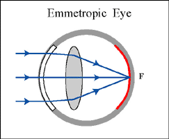
In an emmetropic eye, light rays from far away (thereby crearing parallel light rays) fall on the fovea. These patients do not need glasses when they look far away.
Myopic Eye

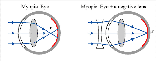
In a myopic eye (=myopia), light rays from far away fall in front of the fovea. To the patient, the image looks blurred. The solution is to provide th patient with a concave ("-") lens.

Note that such a person can look sharp at images close to the eye (like reading) as this will move the focus towards the fovea. That is why these people are called "nearsighted"

Hyperopic Eye

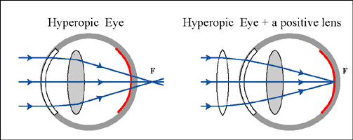
In a hyperopic eye (=hyperopia), parallel light rays will fall "behind" the retina. To help these patients, a convex ("+") lens is required which will help 'break' the light rays more.

Note that these patients can (and do) help themselves by accomodating their lens. This will also move the focus onto the fovea. They do this automatically and therefore often they don't know that they have a refraction anomaly. They are therefore called "farsighted". They will often complain of headaches or tiredness as their ciliary muscle contracts all the time.


G. Astigmatism:

Astigmatism 1:

in the previous refraction anomalies (myopia and hyperopia) the curvature or the bending of the lens was the same at all angles. In some patients, the degree of curvature is different between two (or more) different angles. In the left diagram, this is illustrated by a flat convex lens that is more curved from top to bottom then from left to right. An exagerated example of such a curvature is the surface of an egg; more curved in one direction then in another.

Astigmatism 2:

The consequence of astigmatism is that there is not one focal point but several, located between one extreme and the other. Such patients require specially designed lenses in which the curvature of the lens is adapted to their astigmatism. This can be seen in some lens prescription as follows: +1.5 D at 100 degrees and +0.5 at 45 degrees.


H. Cataract:

A normal eye; the pupil is black.

Cataract:

In some patients, the lens becomes, gradually, non transparant. There are many reasons for this to happen including metabolic diseases, congenital or old age. Because the lens becomes gradually less transparent, the patients will see the images more and more blurred. The therapy is to remove the lens and to replace it with a new artificial intra-ocular lens.

Cataract; the pupil is cloudy or 'milky', 'bluish'

'

Why does the eye (pupil) look red in some flash pictures?


I. Intra-ocular pressure and Glaucoma:

Anterior and Posterior Chamber:

The space between the cornea and the lens is filled with a fluid (= the aqeuous humor). The iris divides this fluid in two spaces; a posterior and an anterior chamber (chamber = room).

Intra-ocular circulation 1:

The ocular fluid is secreted by the ciliary body into the posterior chamber. From there, it flows through the pupil (= the opening between the iris), into the anterior chamber. There, it flows back into the corner between the sclera, the base of the iris and the ciliary body.

Intra-ocular circulation 2:

In this (narrow) angle there are trabeculae that filter the fluid (like a delta) into a canal (= canal of Schlemm) which, in turn, drains into a local vein.

Intra-ocular circulation 3:

In this manner, fresh fluid (with nutrients etc) is constantly flowing through the anterior part of the eye and, by diffusion, through the lens and the vitreous body towards the retina.

Intra-ocular pressure:

This also provides for a small pressure in the eye of about 5-10 mmHg. This keeps the eye in the shape of a ball and all its internal structures (lens etc) straight. If the pressure were too low, then the eyeball would collapse and vision becomes blurred.

Glaucoma 1:

If the pressure gets too high (=glaucoma), then another danger arises. A too high pressure (> 20 mmHg) will impede or block blood flow through the optic nerve. These vessels are crucial as they perfuse the retina.

Glaucoma 2:

If the blood perfusion is stopped, the photoreceptor cells will become ischemic (= no blood) and die. The person will become blind.

Glaucoma 3:

An acute glaucoma (pressures 70-80 mmHg) can occur if there is a sudden obstruction of the flow to the canal of Schlemm. A chronic glaucoma (pressures 20-30 mmHg) occurs when the obstruction is limited.


© HumanPhysiology.academy 2014